
Basic structure
Cross section of a bovine kidney

Figure 1: The cross section of the kidney
Image courtesy of https://www.flickr.com/photos/anicole/3046330947/sizes/o/
This image is under a Creative Commons license.
2 distinct layers are observed. The outer layer is cortex and the inner layer is medulla. The latter has a deeper red colour because of its rich blood supply. The innermost structure is renal pelvis which consists of fat tissues. The renal pyramid can be seen clearly in this picture.
Nephron
Nephrons are the basic functional units of the kidney. Each kidney consisits about 400,000-800,000 nephrons. The number of nephron declines with aging. There are 2 populations of nephrons, cortical/superificial and juxtamedullary nephrons. The difference between these 2 types is the length of the loop of Henle. The loop of juxtamedullary nephron extends into deep inner medulla which plays an important role of concentrating urine.
Blood arrives at the glomerulus / renal corpuscle via afferent arteriole and is filtered in the glomerular capsule. The filtrate is then drained into proximal convoluted tubule (PCT) where most of the reabsorption takes place. It then travels along the loop of Henle where the tubular fluid is concentrated by a process called countercurrent multiplication. Following by the loop, it is distal convoluted tubule (DCT) which loops back to the cortex and pass around the glomerulus.
There are specialised tubular cells in DCT near the glomerulus called macula densa. The smooth muscle cells around the afferent arteriole and the mucula densa together form juxtaglomerular apparatus (JGA). It helps to regulate the blood pressure and the tubular flow within the kidney.
The last part of DCT continues with the collecting duct (CD) where the concentration of urine is tightly regulated. Different collecting ducts join together to form papillary duct which will end at the calyx at the renal pelvis.
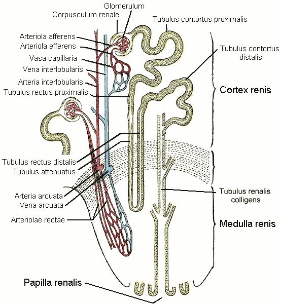
Figure 2: Nephron
Image courtesy of https://commons.wikimedia.org/wiki/File:Kidney_nephron.png
This image is in the public domain.